
Management of Ureteral Stones
Ureteral stone disease is among the most painful and prevalent of urologic disorders. As many as 7% of female Americans and 12% of male Americans will be affected by urinary stones at some point in their lives, and these numbers are rising secondary, likely due to environmental and dietary changes in the modern world.
Fortunately, most patients who are diagnosed with a ureteral stone will be able to pass their stone without any intervention or surgical procedures. However, if you are not so lucky as to pass your stone spontaneously, the following information should help you and your doctor address the causes, symptoms and possible complications created by your ureteral stone disease.

How does the urinary tract work under normal conditions?
The urinary tract is similar to a plumbing system, with a series of special pipes that transport water and salts through the body. A normal urinary tract includes two kidneys, two ureters, the bladder, and the urethra.
The kidneys act as a filter system for the blood, cleansing it of poisonous materials while retaining valuable sugars, salts and minerals. Urine, the waste product of the filtration process, is produced in the kidney and continuously trickles through two 10- to 12-inch long tubes called ureters, all the way from the kidney to the bladder.
The ureters are about one-fourth of an inch in diameter and their muscular walls contract to make waves of movement that forces urine into the bladder. The bladder is an expandable structure, and stores the urine until it can be conveniently disposed. The tube through which the urine flows out of the body is called the urethra.
What is a ureteral stone?
A ureteral stone is a kidney stone that has left the kidney and moved down into the ureter. The stone begins as a tiny grain of undissolved material located where urine collects in the kidney. When the urine flows out of the kidney, this grain of undissolved material is left behind. The material deposited is usually a mineral called calcium oxalate; there are other, less common, materials that may also form a kidney stone such as cystine, calcium phosphate, uric acid and struvite. Over time, more and more undissolved material is deposited, and the stone progressively becomes larger. Most stones enter the ureter when they are still small enough to pass down the entire ureter into the bladder. From there, the stone passes out of the body with urination. Some stones, by the time they leave the kidney, have grown too large to pass through the entire ureter. Such stones may become lodged in a narrow part of the ureter, causing pain and possibly blocking the flow of urine. These stones may need to be treated.
What are the signs of a problem?
Usually, the initial symptom of a kidney stone is one of extreme pain, described by some as being worse than that associated with childbirth. The pain often begins suddenly, when the stone moves within the kidney or ureter and causes irritation or blockage. Typically, a person feels a sharp, cramping pain in the back or side, the region of the body where the kidney is located. In some cases, the pain may radiate to the lower abdomen or groin. Oftentimes, blood in the urine and nausea and vomiting will accompany the pain, too.
There are some situations when stones do not produce any obvious symptoms. However, although the stones may be "silent," they can still be growing, and may even threaten irreversible damage to kidney function. In other cases, a stone may not be large enough to cause major symptoms, but may still trigger a dull ache that is often confused with muscle or intestinal pain. It is important to emphasize, too, that if fever or chills accompany any of these symptoms, then there may be an infection. You should contact your urologist immediately.
If the stone is too large to pass easily, pain continues as the muscles in the wall of the tiny ureter try to squeeze the stone along into the bladder and the kidney stretches from backup of urinary flow. One may feel the need to urinate more often or feel a burning sensation during urination. In a man, pain may move down to the tip of the penis. If the stone is close to the lower end of the ureter at the opening into the bladder, a person will frequently feel like they have not fully completed urination.
Stone size is an important consideration when dealing with a ureteral stone. Although some stones as small as 2mm have caused many symptoms while those as large as a pea have quietly passed, the general rule however is that stones 5 mm or less are more likely to pass without the need for intervention and stones larger than 5 mm are unlikely to pass spontaneously.
How are ureteral stones diagnosed?
Sometimes "silent" stones ? those that cause no symptoms ? are found on X-rays taken during a general health exam. These stones would likely pass unnoticed. If these incidentally detected stones are large, then treatment may be considered. More often, though, ureteral stones are found on imaging obtained when someone who complains of blood in the urine or sudden pain. These diagnostic images give the doctor valuable information about the stone's size and location. Blood and urine tests can also help detect any abnormalities that might complicated the management of the stone.
If your doctor suspects a stone but is unable to make a diagnosis from a simple X-ray, he or she may scan the urinary system with computed tomography (CT) or intravenous pyelography (IVP). IVP is an imaging technique that utilizes radiopaque injections of dye followed, during excretion by the kidneys, by abdominal X-rays. A kidney obstructed by a stone will not be able to excrete the dye as quickly and may also appear enlarged when compared to the normal, unobstructed kidney. In many hospitals the IVP has been replaced by a CT scan, which is an extremely rapid and accurate diagnostic tool, which will detect almost all types of ureteral stones.
What are some treatment options?
Treating kidney stone disease depends largely on the size, position and number of stones that are present. Luckily, the majority of small stones (5 mm in diameter or less) will pass if you simply drink plenty of fluids each day. Consuming two to three quarts of water increases urine production, which eventually washes the stones out of the system. Once a stone has passed, no other treatment is necessary. It is always helpful to capture the passed stone, if possible, so that it may be analyzed to see what it is made of. Recent studies suggest that the majority of stones (95 percent) that are capable of spontaneous passage will pass within six weeks. After that time, continued observation is probably not warranted, and if the stone has not passed, further treatment will likely be needed.
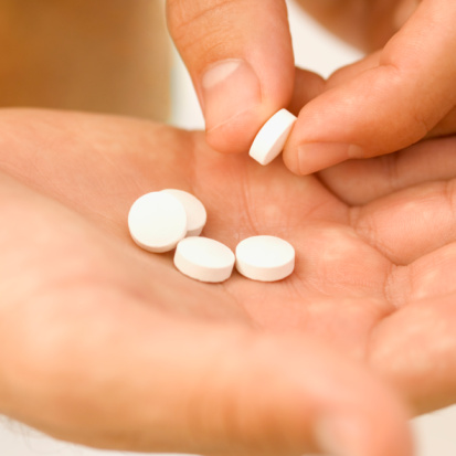
The sudden pain that occurs when small stones start to move down the ureter can usually be treated with hydration and painkillers (anti-inflammatories and narcotics). Some people may also need anti-nausea medications. Certain types of stones, such as those made of uric acid, can be dissolved with medical therapy. The majority, however, are composed of calcium and will not be dissolved with medicine alone.
Surgery should be reserved as an option for cases where more conservative approaches have failed. Surgery may be needed if a stone:
- does not pass after a reasonable period of time
- causes constant and intractable pain
- Causes persistent nausea and vomiting, such that the patient is not able to tolerate food or liquid
- is too large to pass on its own
- blocks the flow of urine
- causes ongoing urinary tract infection
- harms the function of the kidney
Historically, the surgical removal of a kidney stone involved an operation with an incision and an often lengthy recovery time. Today, though, most stones can be treated in a minimally invasive, or even non-invasive, fashion. As a result, recovery times are now measured in days, not weeks. Some of the treatments for kidney stones include:
Extracorporeal shock wave lithotripsy (ESWL ®). ESWL® Is the most frequently used procedure for eliminating kidney stones. Shock wave treatment uses a machine called a lithotripter, which works by directing ultrasonic or shock waves, created outside your body ("extracorporeal") through skin and tissue, until they hit the dense kidney stones. The impact of the shock wave causes stress on the stone; the cumulative effect of repeated shock waves is one of increasing stress on the stone, until eventually the stone crumbles into small pieces. These small pieces, about the size of grains of sand, usually pass easily through the urinary tract, and are voided out in the patient?s urine. Shock wave lithotripsy is generally used when the stone is not excessively large, the kidney is functioning well, and there is no blockage to the passage of stone fragments.
In the original ESWL devices, the patient used to recline in a water bath while the shock waves were transmitted. Today, the machines are more compact and have a soft cushion on which the patient lies. Ultrasound or fluoroscopy is used to locate the stone and focus the shock waves.
In most cases, shock wave lithotripsy is done on an outpatient basis. Recovery time is short and most people can resume normal activities in a few days. However, one ESWL® session by itself may not free the patient of all stone material. A repeat ESWL® session may be necessary. ESWL® is not the ideal treatment choice for all patients. Patients who are pregnant, obese, have obstruction past the stone, have abdominal aortic aneurysms, urinary tract infections or uncorrected bleeding disorders should not have ESWL®. In addition, certain factors such as stone size, location and composition may necessitate other alternatives for stone removal.
Because of possible discomfort during the procedure, anesthesia or sedation is generally needed. Although ESWL can be performed with heavy sedation alone, success rates may be better when a general anesthetic is used. Once the treatment is completed, the small stone particles then pass down the ureter and are eventually urinated away. In certain cases, a stent may need to be placed up the ureter prior to SWL to assist in locating the stone or prevent the kidney from being obstructed by passage of stone fragments.
Certain types of stone (cystine, calcium oxalate monohydrate) can be resistant to SWL and may necessitate an alternative treatment approach.. In addition, larger stones may not break up into pieces small enough to be discharged from the kidney. Stones located in the lower portion of the kidney also have a decreased chance of passage.
While shock wave lithotripsy is considered safe and effective, it can still cause complications. Most patients have blood in their urine for a few days after treatment. Bruising and minor discomfort in the back or abdomen from the shock waves are also common. To reduce the risk of complications, urologists usually tell their patients to avoid aspirin and other drugs that affect blood clotting for a period of time before treatment. Another complication may occur if the stone fragments cause discomfort as they pass through the urinary tract. In some cases, the urologist will insert a small tube called a stent through the bladder prevent this complication.
Ureteroscopy (URS). Ureteroscopy involves the use of ureteroscopes, small flexible or semi-rigid telescopes that can be inserted up the urethra, through the bladder and into the ureter without an incision. These instruments allow the doctor to view a ureteral stone directly. They also have small working channels through which various devices can be passed to remove or fragment the stone. Anesthesia is generally used, and a stent is generally left in the ureter for a few days after treatment while healing takes place. Ureteroscopy was developed in the 1970s and came into wide use during the 1980s and 1990s. The risks of ureteroscopy include perforation or stricture (scar tissue), especially if the stone has been impacted or embedded within the wall of the ureter for longer than two months. The majority of ureteroscopic procedures can be performed as day surgery and most individuals can return to work within several days following the procedure.
Percutaneous nephrolithotomy (PNL). This procedure is the treatment of choice for patients with ureteral stones that are larger, are in a location that does not allow effective use of SWL, or cause a blockage so severe that they cannot be bypassed using stent.
In this procedure, the surgeon makes a tiny cut in the flank area and then uses an instrument called a nephroscope to locate and remove the stone. For larger stones, a type of energy probe (ultrasonic, electrohydraulic, hydraulic, laser, or pneumatic) may be needed to break the stone into small pieces. All of this is done while the patient is sedated or under anesthesia.
One advantage of this procedure over SWL is that the surgeon removes the stone fragments instead of relying on their natural passage from the ureters. Generally, patients stay in the hospital overnight and may have a small catheter in the kidney and/or stent during the healing process. Most patients can resume light activity in one to two weeks.

Open surgery. This is the most invasive treatment and is rarely performed these days. In open surgery for ureteral stones, the doctor makes a surgical cut to expose the ureter where the stone is located. Another cut is made in the ureter itself, and the stone is directly removed. Open surgery is reserved for highly complicated, difficult cases. Most patients need about six weeks to recover after the operation.
What can be expected after treatment?
Although stone recurrence rates differ with individuals, in general you have a 50 percent chance of redeveloping stones within the next five years. So prevention is essential. Your urologist may follow-up with several tests to determine which factors (e.g., medication or diet) should be changed to reduce your risk. Treatment results can vary depending on the selected treatment approach, as well as patient and stone specific factors.
Content provided courtesy & permission of the American
Urological Association Foundation, and is current as of 5/2010.
Visit us at www.urologyhealth.org for additional information.